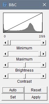
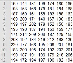
What is a Raster Image?
- Raster images are composed of a grid of pixels
- Each pixel contains intensity information
- Number of bits (N) determine the range of intensity levels
| Image Type | Range of intensity levels (0 to 2N-1) |
|---|---|
| 8-bit | 0-255 |
| 16-bit | 0-4095 |
| 32-bit | 0-65,535 |
| RGB color (3 x 8 bits) | 0-255 per channel |
Segmentation using global thresholding
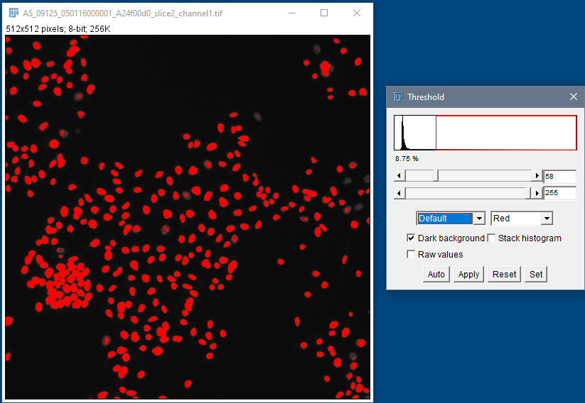
Human HT29 colon cancer cells, Image from Broad Bioimage Benchmark Collection, Ljosa et al. 2012 Nat Methods
Segmentation using global thresholding
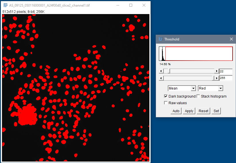
Human HT29 colon cancer cells, Image from Broad Bioimage Benchmark Collection, Ljosa et al. 2012 Nat Methods
Segmentation with local thresholding
Auto Local
Threshold
in Fiji
![]()
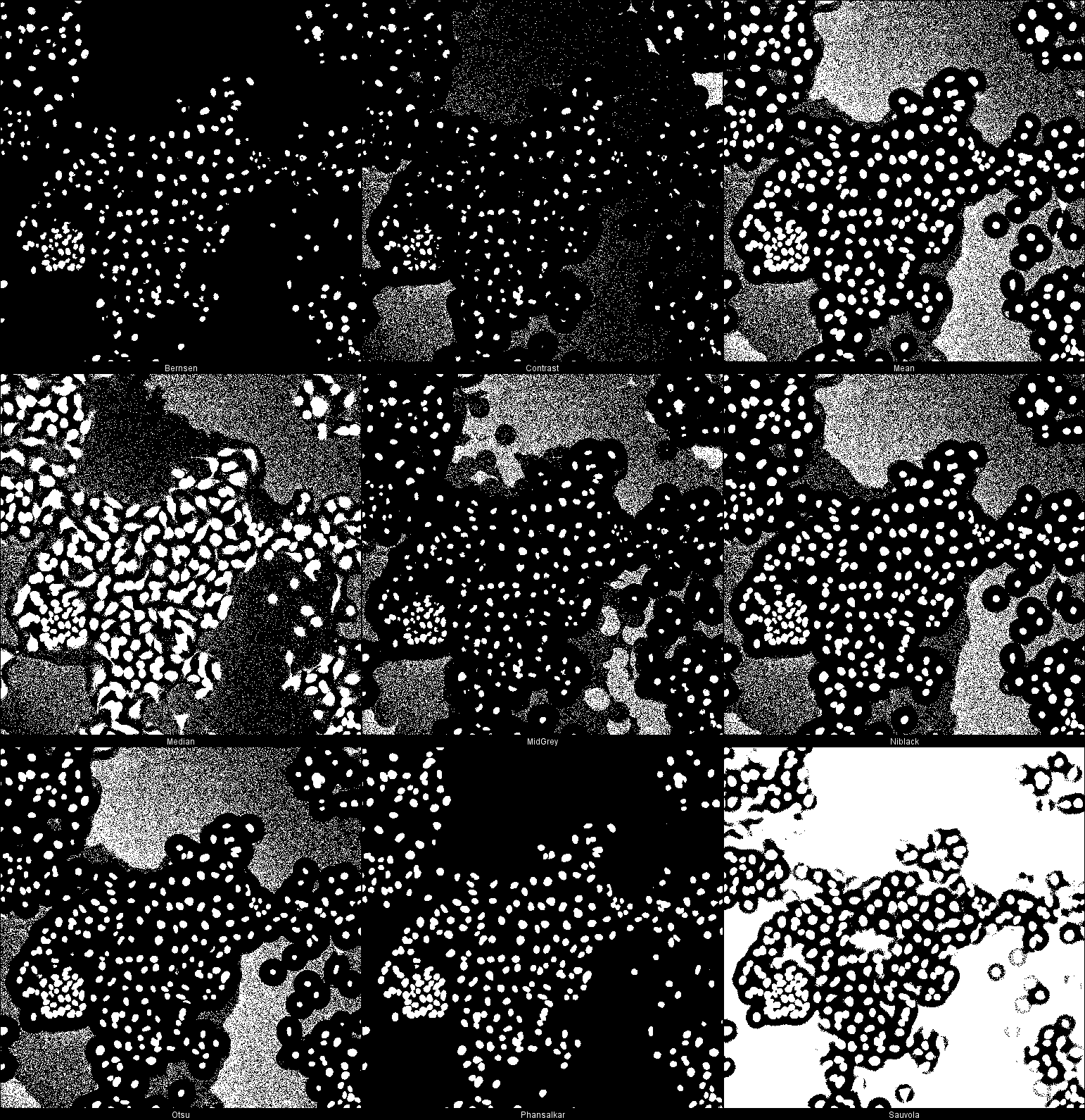
Human HT29 colon cancer cells, Image from Broad
Bioimage Benchmark Collection, Ljosa et al. 2012 Nat Methods
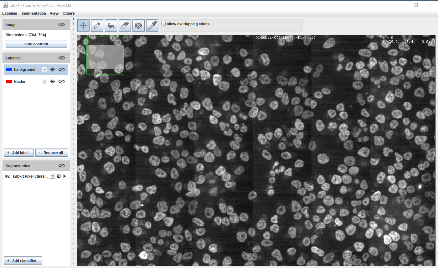
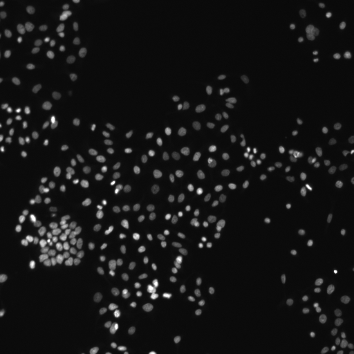
Hoechst-stained Nuclei, image courtesy of Cherie Au, Giannakakou Lab, Weill Cornell Medicine
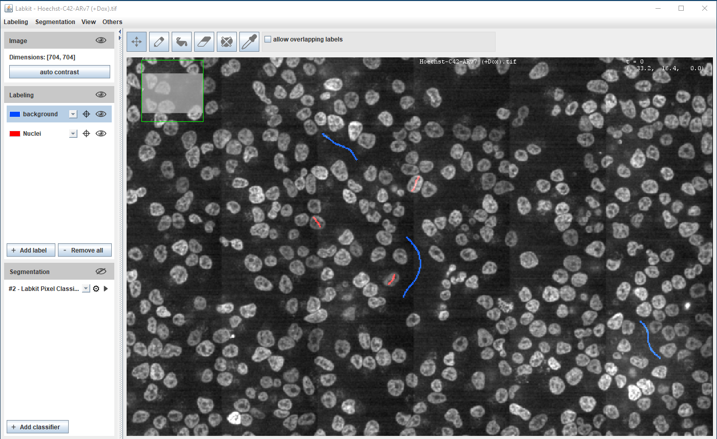
Hoechst-stained Nuclei, image courtesy of Cherie Au, Giannakakou Lab, Weill Cornell Medicine
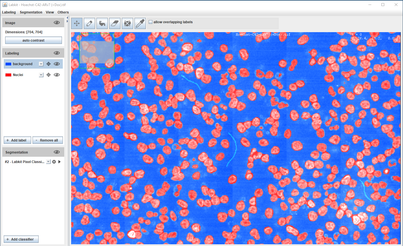
Hoechst-stained Nuclei, image courtesy of Cherie Au, Giannakakou Lab, Weill Cornell Medicine
Segmentation using Deep Learning
Most accurate methods available for cells/nuclei segmentation
Step 1: Training
Generating a Deep Learning model is resource hungry:
- High-end workstation
- Large amounts of training data (images and annotations)
- Training could take hours to days
- Good programming knowledge required - Python
Step 2: Prediction
Using the model from step 1 to predict the segmentation results :
- A regular laptop is just fine
- Prediction takes seconds to mins
- Little to no programming knowledge required
Segmentation using Deep Learning
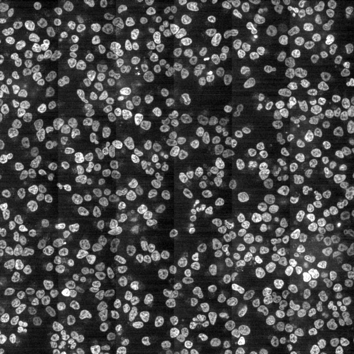
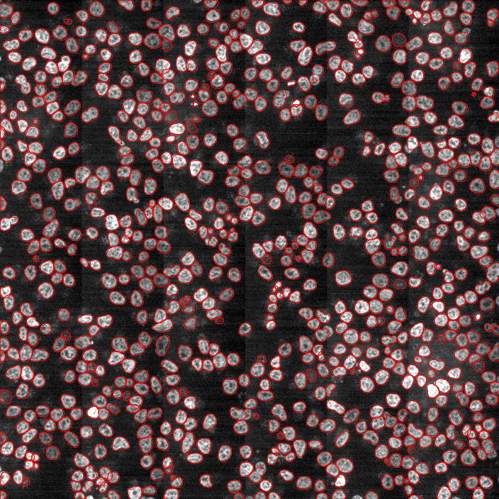
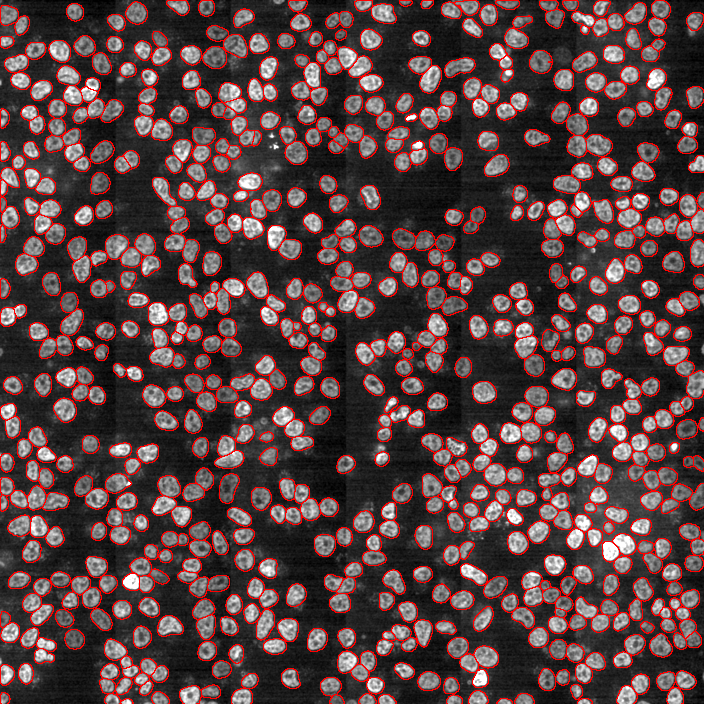
Hoechst-stained Nuclei, image courtesy of Cherie Au, Giannakakou Lab, Weill Cornell Medicine
Workshop Exercise 1: StarDist based nuclear segmenation in a challenging image in Fiji
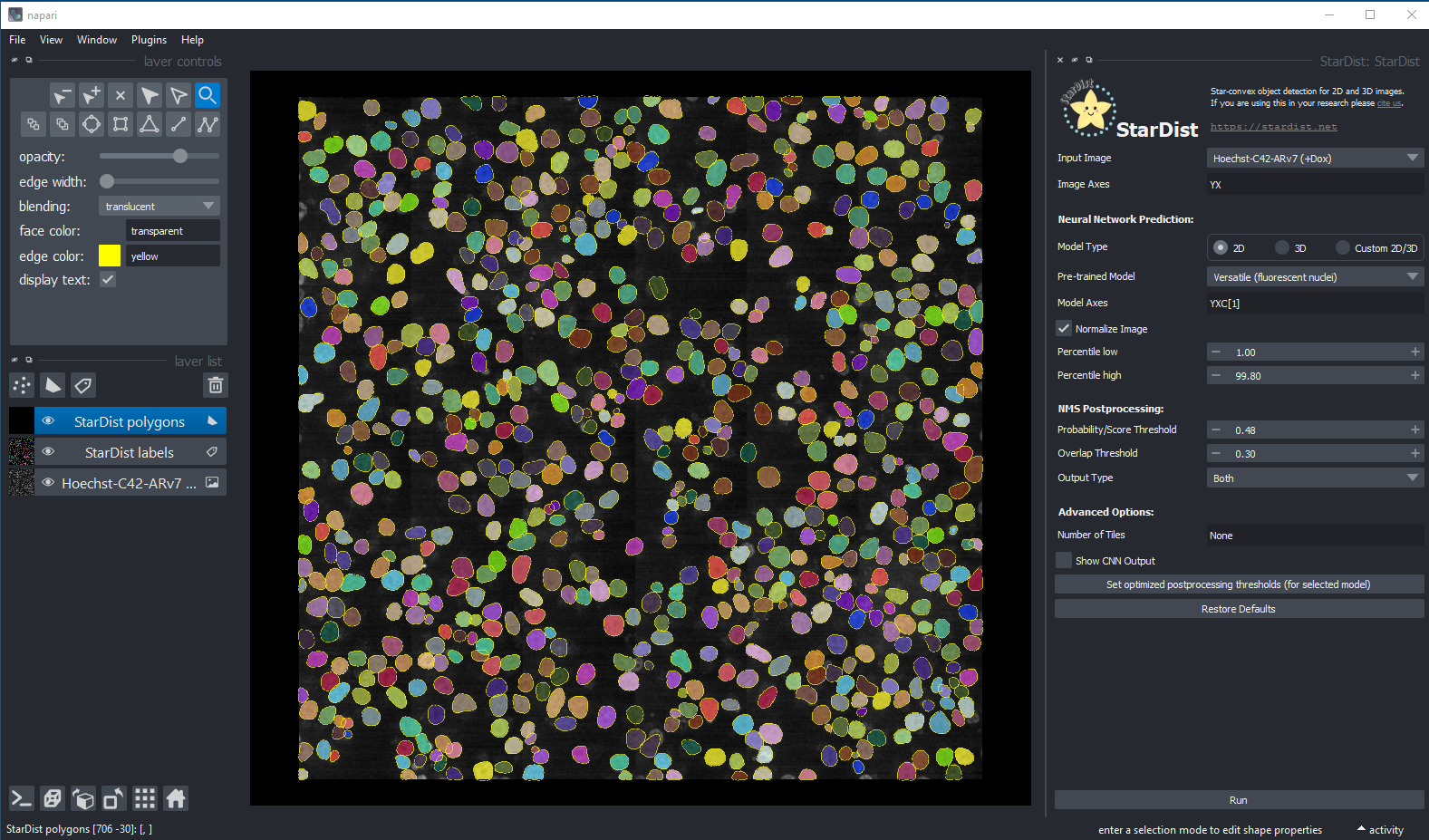
Segmentation using StarDist in Napari
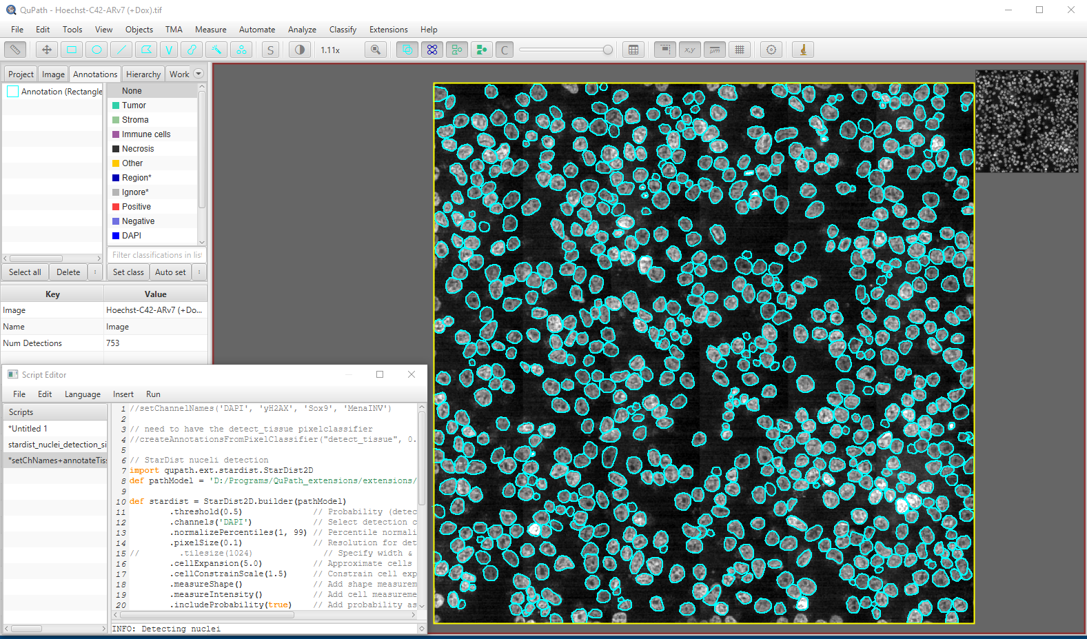
Segmentation using StarDist in Qupath
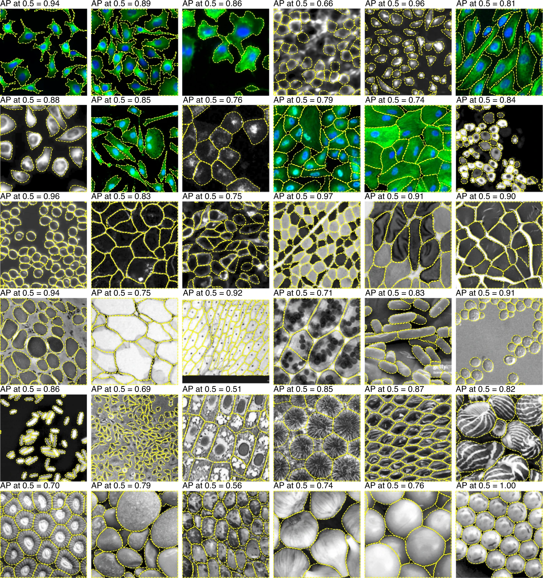
Cellpose
Stringer et al 2021, Nat Methods
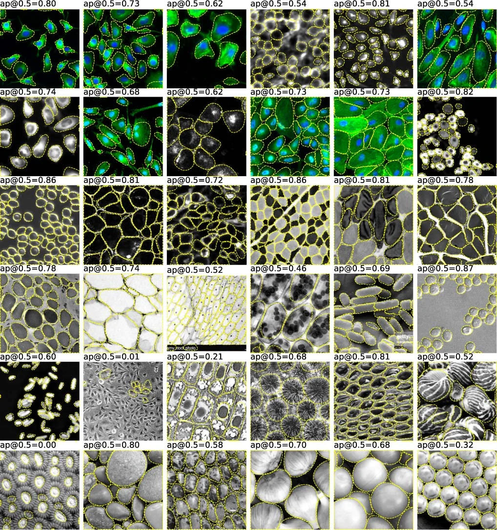
StarDist
Stringer et al 2021, Nat Methods
1. Cell crowding

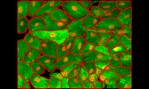
Image from https://github.com/MouseLand/cellpose
2. Cell crowding + noisy signal
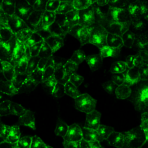
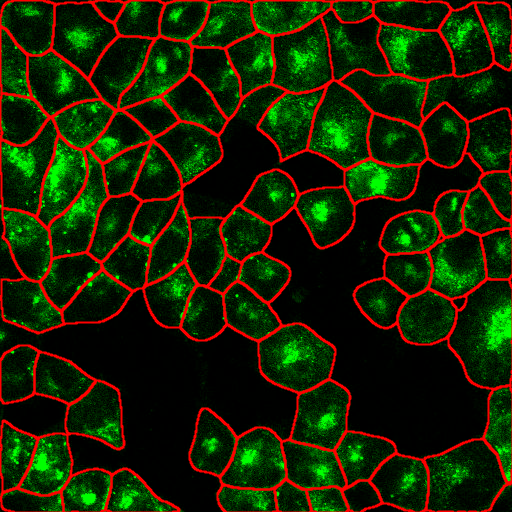
Image from https://github.com/MouseLand/cellpose
3. Cell crowding + uneven illumination
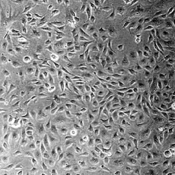
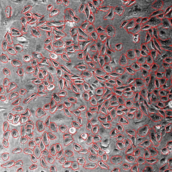
Image from https://github.com/MouseLand/cellpose